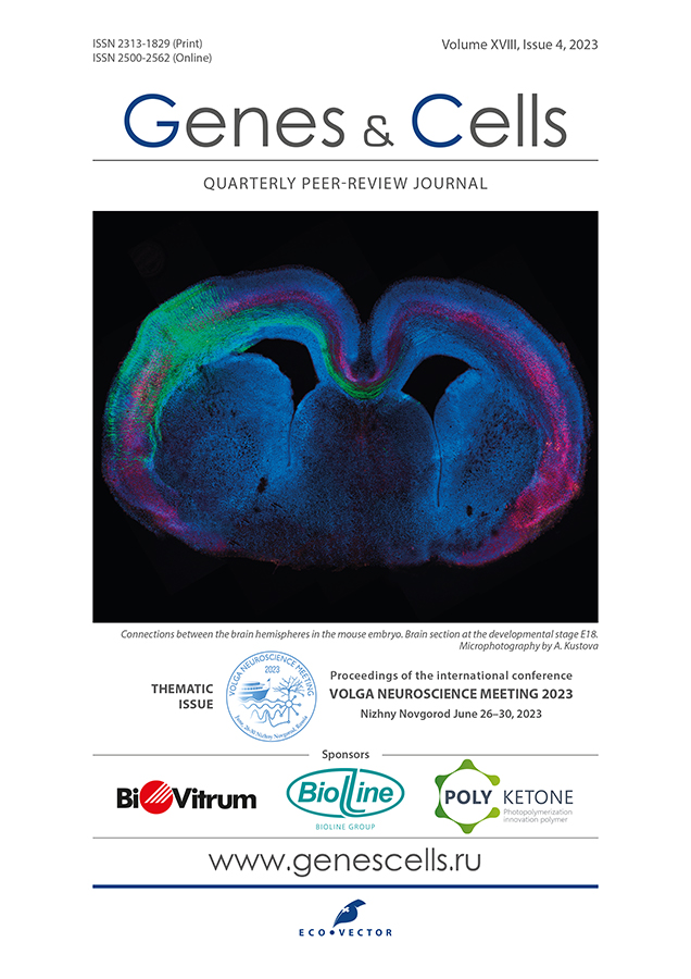
Developmental expression patterns of genes mutated in patients with neurodevelopmental disorders
- Authors: Kondakova E.V.1,2, Gavrish M.S.1, Filat’eva A.E.1, Tarabykin V.S.3
-
Affiliations:
- Institute of Neurosciences, National Research Lobachevsky State University of Nizhny Novgorod
- Research Institute of Medical Genetics, Tomsk National Research Medical Center of the Russian Academy of Sciences
- Institute of Cell Biology and Neurobiology, Charité-Universitätsmedizin Berlin
- Issue: Vol 18, No 4 (2023)
- Pages: 494-497
- Section: Conference proceedings
- Submitted: 16.11.2023
- Accepted: 17.11.2023
- Published: 15.12.2023
- URL: https://genescells.ru/2313-1829/article/view/623462
- DOI: https://doi.org/10.17816/gc623462
- ID: 623462
Cite item
Abstract
Neurodevelopmental disorders (NDDs) comprise a heterogeneous spectrum of disorders with diverse manifestations, such as microcephaly, structural brain abnormalities, epilepsy, developmental delay, intellectual disability, and autism spectrum disorders [1]. Although relatively rare, each type of NDDs represents a significant population of neurological patients. The global prevalence of NDDs exceeds 15% [2]. NDDs typically arise from molecular cascades that are highly regulated and disrupted by either gene mutations or environmental factors. The genetic basis of a substantial proportion of such disorders is hard to discern given that not all are inherited according to Mendelian principles and involve allelic variations from multiple genes. However, roughly 40% of NDDs are believed to be caused by the disruption of a single gene, indicating monogenic conditions. [3]
Understanding and predicting the physiological function of a protein encoded by a specific gene, determining its interactions with other proteins, and investigating the role of the gene in organ and tissue development requires a close examination of its expression. Thus, a crucial initial step in researching genes associated with neurodevelopmental disorders is to investigate the expression patterns of these genes in the mouse brain at various embryonic developmental stages.
In situ RNA hybridization was used to analyze gene expression patterns in slices of mouse brain tissue. Fixed in 4% paraformaldehyde/phosphate-buffered saline/diethylpyrocarbonate mouse brain samples at embryonic (E12.5, E15.5, E18.5) and postnatal (P1, P21) developmental stages were sectioned using a Leica CM1520 cryostat with 15 µm slice thickness. Next, we conducted in situ hybridization of cellular mRNA using DIG-dUTP-labeled RNA probes that were previously synthesized by PCR with cDNA and gene-specific primers. 5-bromo-4-chloro-3-indolyl phosphate/nitroblue tetrazolium (BCIP/NBT) was used, which produces an insoluble dark blue or purple sediment visible under a light microscope by reaction with alkaline phosphatase, to visualize the localization of mRNA expression in tissues.
The study examined the expression of a member of the CCDC gene family which encodes proteins involved in intercellular transmembrane signal transduction. In situ hybridization was performed on mouse brain slices, revealing significant mRNA expression of the gene in the cerebral cortex. Additionally, mouse knockout experiments are planned to investigate the gene’s role in brain development.
Full Text
Neurodevelopmental disorders (NDDs) comprise a heterogeneous spectrum of disorders with diverse manifestations, such as microcephaly, structural brain abnormalities, epilepsy, developmental delay, intellectual disability, and autism spectrum disorders [1]. Although relatively rare, each type of NDDs represents a significant population of neurological patients. The global prevalence of NDDs exceeds 15% [2]. NDDs typically arise from molecular cascades that are highly regulated and disrupted by either gene mutations or environmental factors. The genetic basis of a substantial proportion of such disorders is hard to discern given that not all are inherited according to Mendelian principles and involve allelic variations from multiple genes. However, roughly 40% of NDDs are believed to be caused by the disruption of a single gene, indicating monogenic conditions. [3]
Understanding and predicting the physiological function of a protein encoded by a specific gene, determining its interactions with other proteins, and investigating the role of the gene in organ and tissue development requires a close examination of its expression. Thus, a crucial initial step in researching genes associated with neurodevelopmental disorders is to investigate the expression patterns of these genes in the mouse brain at various embryonic developmental stages.
In situ RNA hybridization was used to analyze gene expression patterns in slices of mouse brain tissue. Fixed in 4% paraformaldehyde/phosphate-buffered saline/diethylpyrocarbonate mouse brain samples at embryonic (E12.5, E15.5, E18.5) and postnatal (P1, P21) developmental stages were sectioned using a Leica CM1520 cryostat with 15 µm slice thickness. Next, we conducted in situ hybridization of cellular mRNA using DIG-dUTP-labeled RNA probes that were previously synthesized by PCR with cDNA and gene-specific primers. 5-bromo-4-chloro-3-indolyl phosphate/nitroblue tetrazolium (BCIP/NBT) was used, which produces an insoluble dark blue or purple sediment visible under a light microscope by reaction with alkaline phosphatase, to visualize the localization of mRNA expression in tissues.
The study examined the expression of a member of the CCDC gene family which encodes proteins involved in intercellular transmembrane signal transduction. In situ hybridization was performed on mouse brain slices, revealing significant mRNA expression of the gene in the cerebral cortex. Additionally, mouse knockout experiments are planned to investigate the gene’s role in brain development.
ADDITIONAL INFORMATION
Funding sources. The study was funded by the Ministry of Science and Higher Education of the Russian Federation (grant No. FSWR-2023-0029).
Authors' contribution. All authors made a substantial contribution to the conception of the work, acquisition, analysis, interpretation of data for the work, drafting and revising the work, and final approval of the version to be published and agree to be accountable for all aspects of the work.
Competing interests. The authors declare that they have no competing interests.
About the authors
E. V. Kondakova
Institute of Neurosciences, National Research Lobachevsky State University of Nizhny Novgorod; Research Institute of Medical Genetics, Tomsk National Research Medical Center of the Russian Academy of Sciences
Author for correspondence.
Email: elen_kondakova@list.ru
Russian Federation, Nizhny Novgorod; Tomsk
M. S. Gavrish
Institute of Neurosciences, National Research Lobachevsky State University of Nizhny Novgorod
Email: elen_kondakova@list.ru
Russian Federation, Nizhny Novgorod
A. E. Filat’eva
Institute of Neurosciences, National Research Lobachevsky State University of Nizhny Novgorod
Email: elen_kondakova@list.ru
Russian Federation, Nizhny Novgorod
V. S. Tarabykin
Institute of Cell Biology and Neurobiology, Charité-Universitätsmedizin Berlin
Email: elen_kondakova@list.ru
Germany, Berlin
References
- Mitani T, Isikay S, Gezdirici A, et al. High prevalence of multilocus pathogenic variation in neurod velopmental disorders in the Turkish population. American Journal of Human Genetics. 2021;108(10):1981–2005. doi: 10.1016/j.ajhg.2021.08.009
- Received: 19.06.2023 Accepted: 26.11.2023 Published online: 20.01.2024
- Barkovich AJ, Guerrini R, Kuzniecky RI, et al. A developmental and genetic classification for malformations of cortical development: update 2012. Brain. 2012;135(Pt 5):1348–1369. doi: 10.1093/brain/aws019
- Mesnil M, Defamie N, Naus C, Sarrouilhe D. Brain Disorders and Chemical Pollutants: A Gap Junction Link? Biomolecules. 2020;11(1):51. doi: 10.3390/biom11010051
Supplementary files


