

")


Soluble factors formed during the healing of the endometrium suppress its "fibrosis” in vitro
- Authors: Eremichev R.Y1, Grigorieva O.A1, Kulebyakin K.Y1, Efimenko A.Y.1, Makarevich P.I1
-
Affiliations:
- Institute of Regenerative Medicine, Medical Research Centre of M.V. Lomonosov Moscow State University
- Issue: Vol 13, No 2 (2018)
- Pages: 63-66
- Section: Articles
- Submitted: 05.01.2023
- Published: 15.06.2018
- URL: https://genescells.ru/2313-1829/article/view/120738
- DOI: https://doi.org/10.23868/201808021
- ID: 120738

Cite item

Abstract
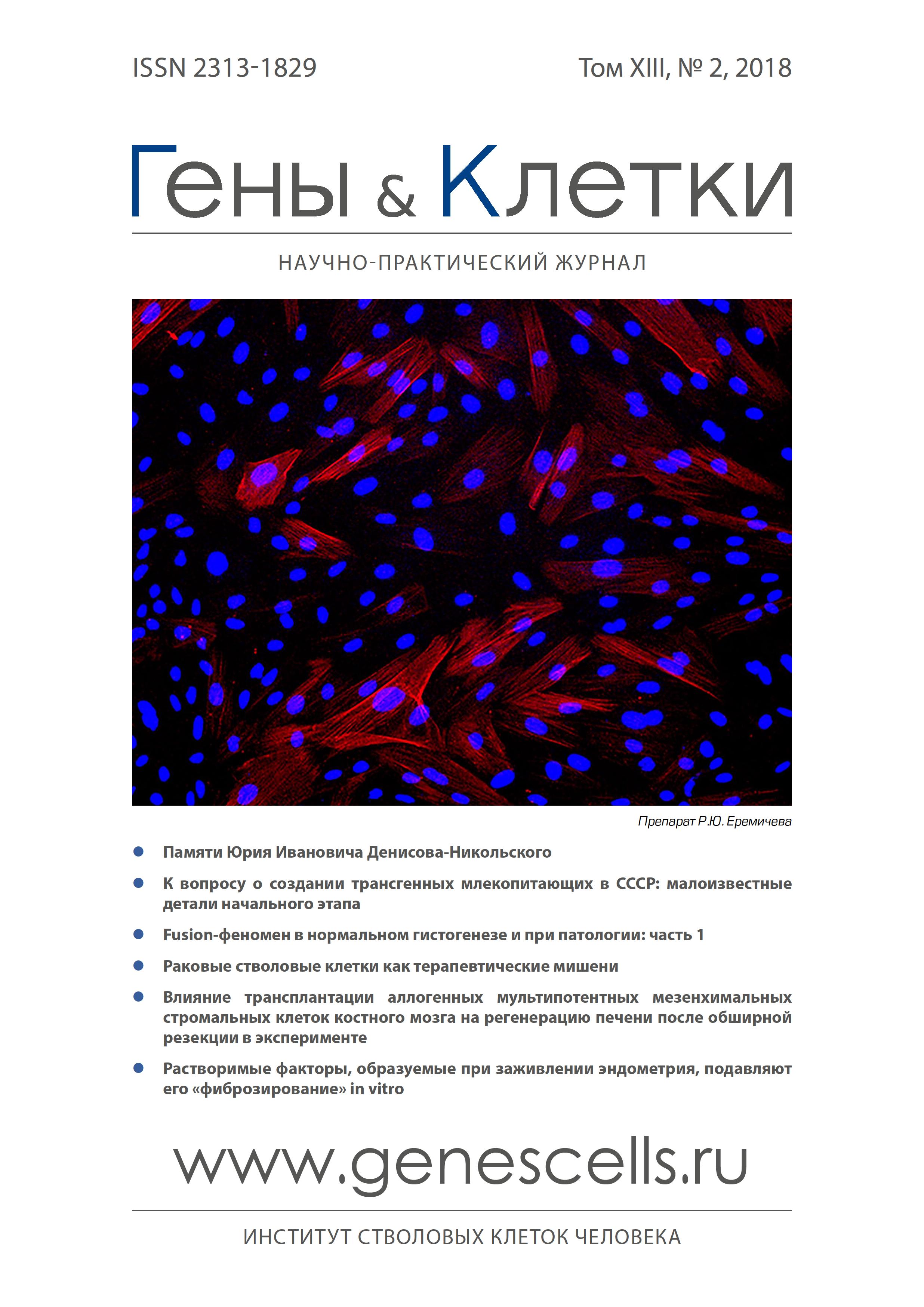
During each period, the uterine mucosa of women of reproductive age heals without fibrosis. Previously, we established that the soluble factors that are released in this way have an antifibrotic effect on the culture of the human endometrial mesenchymal stromal cells. The objective of this work was to evaluate the antifibrotic properties of these factors on the in vitro endometrial fibrosis model. Serum menstrual and peripheral blood were obtained from a healthy donor in one day. Mesenchymal stromal cells of the endometrium were also isolated from menstrual blood. Simulation of endometrial fibrosis in vitro was carried out by differentiation of endometrial mesenchymal stromal cells into myofibroblasts under the action of TGF-ß1 (5 ng/ml). Evaluation of the effectiveness of the menstrual blood serum antifibrotic effect on the endometrial mesenchymal stromal cells and myofibroblasts derived from them was carried out by analyzing the expression of а-smooth muscle actin by immunofluorescence. Serum of peripheral blood with equal protein concentration was used as a control. Menstrual blood serum reduces the number of stress-fibrils positive for а-smooth muscle actin (a marker of myofibroblasts), both in the culture of endometrial mesenchymal stromal cells, and in in vitro modeling of endometrial fibrosis using TGF-ß1. These results indicate the presence of soluble factors in the serum of menstrual blood with antifibrotic properties. Perhaps their identification will explain the mechanisms of endometrial healing not accompanied by fibrosis. In addition, it can help to identify the causes of fibrosis of the uterine lining in gynecological diseases and develop effective methods for their treatment.
Full Text
About the authors
R. Y Eremichev
Institute of Regenerative Medicine, Medical Research Centre of M.V. Lomonosov Moscow State University
O. A Grigorieva
Institute of Regenerative Medicine, Medical Research Centre of M.V. Lomonosov Moscow State University
K. Y Kulebyakin
Institute of Regenerative Medicine, Medical Research Centre of M.V. Lomonosov Moscow State University
A. Yu Efimenko
Institute of Regenerative Medicine, Medical Research Centre of M.V. Lomonosov Moscow State University
P. I Makarevich
Institute of Regenerative Medicine, Medical Research Centre of M.V. Lomonosov Moscow State University
Email: pavel.makarevich@gmail.com
References
- R., Hart R., Karthigasu K.A. et al. A re-appraisal of the morphological changes within the endometrium during menstruation: A hysteroscopic, histological and scanning electron microscopic study. Hum Reprod. 2009; 24(6): 1393-401.
- Zhou Q., Wu X., Dai X. et al. The different dosages of estrogen affect endometrial fibrosis and receptivity, but not sdf-1/cxcr4 axis in the treatment of intrauterine adhesions. Gynecol Endocrinol. 2018; 34(1): 49-55.
- Zhu H.Y., Ge T.X., Pan Y.B. et al. Advanced role of hippo signaling in endometrial fibrosis: Implications for intrauterine adhesion. Chin Med J (Engl). 2017; 130(22): 2732-7.
- Shen M., Liu X., Zhang H. et al. Transforming growth factor beta1 signaling coincides with epithelial-mesenchymal transition and fibroblast-tomyofibroblast transdifferentiation in the development of adenomyosis in mice. Hum Reprod. 2016; 31(2): 355-69.
- Ibrahim M.G., Sillem M., Plendl J. et al. Myofibroblasts are evidence of chronic tissue microtrauma at the endometrial-myometrial junctional zone in uteri with adenomyosis. Reprod Sci. 2017; 24(10): 1410-1418.
- Li J., Du S., Sheng X. et al. Microrna-29b inhibits endometrial fibrosis by regulating the sp1-tgf-beta1/smad-ctgf axis in a rat model. Reprod Sci. 2016; 23(3): 386-94.
- Li J., Cen B., Chen S. et al. Microrna-29b inhibits tgf-beta1-induced fibrosis via regulation of the tgf-beta1/smad pathway in primary human endometrial stromal cells. Mol Med Rep. 2016; 13(5): 4229-37.
- Falke L.L., Gholizadeh S., Goldschmeding R. et al. Diverse origins of the myofibroblast-implications for kidney fibrosis. Nat Rev Nephrol. 2015; 11(4): 233-44.
- Pellicoro A., Ramachandran P., Iredale J.P. et al. Liver fibrosis and repair: Immune regulation of wound healing in a solid organ. Nat Rev Immunol. 2014; 14(3): 181-94.
- Kong P., Christia P.,Frangogiannis N.G. The pathogenesis of cardiac fibrosis. Cell Mol Life Sci. 2014; 71(4): 549-74.
- Wynn T.A.,Ramalingam T.R. Mechanisms of fibrosis: Therapeutic translation for fibrotic disease. Nat Med. 2012; 18(7): 1028-40.
- Еремичев Р.Ю. М.О.А., Александрушкина Н.А., Кулебякин К.Ю., Дыйканов Д.Т., Макаревич П.И. Сыворотка менструальной крови оказывает противофиброзное действие на мезенхимные стромальные клетки эндометрия человека. Цитология 2018; 60(2): 96-103.
- Ferguson M.W.,O'Kane S. Scar-free healing: From embryonic mechanisms to adult therapeutic intervention. Philos Trans R Soc Lond B Biol Sci. 2004; 359(1445): 839-50.
- Chen C.Z., Peng Y.X., Wang Z.B. et al. The scar-in-a-jar: Studying potential antifibrotic compounds from the epigenetic to extracellular level in a single well. Br J Pharmacol. 2009; 158(5): 1196-209.
- Talele N.P., Fradette J., Davies J.E. et al. Expression of alpha-smooth muscle actin determines the fate of mesenchymal stromal cells. Stem Cell Reports. 2015; 4(6): 1016-30.
- Anders H.J.,Schaefer L. Beyond tissue injury-damage-associated molecular patterns, toll-like receptors, and inflammasomes also drive regeneration and fibrosis. J Am Soc Nephrol. 2014; 25(7): 1387-400.
- Desai V.D., Hsia H.C.,Schwarzbauer J.E. Reversible modulation of myofibroblast differentiation in adipose-derived mesenchymal stem cells. PLoS One. 2014; 9(1): e86865.
- Yang X., Chen B., Liu T. et al. Reversal of myofibroblast differentiation: A review. Eur J Pharmacol. 2014; 734: 83-90.
Supplementary files
