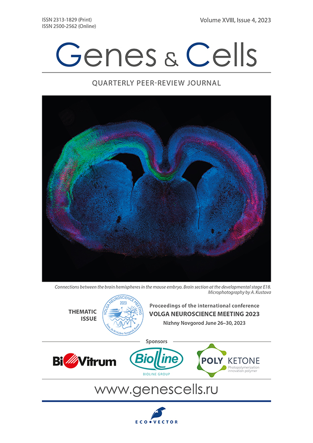
Study of the role of evolutionary new enhancers in the development of the corpus callosum
- Authors: Kustova A.O.1,2, Celis Suescun J.C.1, Rybakova V.P.1,2, Tarabykin V.S.3
-
Affiliations:
- Institute of Neurosciences, National Research Lobachevsky State University of Nizhny Novgorod
- Research Institute of Medical Genetics, Tomsk National Research Medical Center of the Russian Academy of Sciences
- Institute of Cell Biology and Neurobiology, Charité-Universitätsmedizin Berlin
- Issue: Vol 18, No 4 (2023)
- Pages: 502-503
- Section: Conference proceedings
- URL: https://genescells.ru/2313-1829/article/view/623464
- DOI: https://doi.org/10.17816/gc623464
- ID: 623464
Cite item
Abstract
One important aspect of the mammalian brain is the exchange of information between neurons located in different hemispheres. This process evolved during the development of mammals. In marsupial (Marsupialia) and monotreme (Monotremata) mammals, the communication between hemispheres is facilitated through an enlarged anterior commissure. In placental (Eutheria) mammals, a new brain structure, the corpus callosum, emerged during the evolutionary process. The corpus callosum, comprising 80% of the brain’s commissural axons, is the largest commissure in the human body. The corpus callosum is a major contributor to the efficiency of higher neural activities, including memory, decision making, social interaction, and language. A possible explanation for the emergence of a novel structure for interhemispheric interaction is a change in the growth direction of neocortical axons during development. Changes in gene expression levels can regulate this process, which involves controlling axon growth and cell migration in the neocortex. A full comprehension of this process will enable the creation of new animal models for studying cortical malformations and the navigation of axons in the cerebral cortex leading to the formation of interhemispheric connections.
Several enhancers were identified for protein-coding genes with differing expression patterns in neocortical cells of placental and non-placental (marsupial) mammals. Throughout the genomes of the house opossum (Monodelphis domestica) and the house mouse (Mus musculus), the acetylation levels of histone H3 on lysine 27 (H3K27ac) were compared. H3K27ac is considered an epigenetic marker for active gene enhancers. Then, a screening of candidate genes was performed to evaluate their localization and expression levels in the cortex during embryonic development. Thus, the Tbr1 gene was identified. Incorrect expression of this gene may result in changes to cortical development.
The CRISPR/Cas9 system was combined with in utero electroporation to completely delete the active Tbr1 gene enhancer in developing neocortical cells of mouse embryos at day 14 of embryonic development. The impact of this enhancer deletion was then analyzed on the expression of Tbr1 in the upper layers of the cortex, as well as the direction of axon growth and neuronal migration on the 18th day of embryonic development.
A significant reduction in Tbr1 expression was observed in the upper layers of the cortex after deletion of the active enhancer. Only 30% of the electroporated neurons retained Tbr1 expression. Moreover, a considerable delay in neuronal migration was observed in the subventricular zone (41% versus 17% in the control group) and in the upper layers of the cortex (20% versus 35% in the control group). However, the direction of axonal growth remained unchanged: callosal axons effectively crossed the midline and created the corpus callosum.
Thus, expression of the evolutionary novel Tbr1 enhancer is important for neuronal migration during corticogenesis. However, its contribution to the development of the corpus callosum is not fully understood. A detailed analysis of the corpus callosum morphology post-enhancer deletion will be the subsequent step.
Keywords
Full Text
One important aspect of the mammalian brain is the exchange of information between neurons located in different hemispheres. This process evolved during the development of mammals. In marsupial (Marsupialia) and monotreme (Monotremata) mammals, the communication between hemispheres is facilitated through an enlarged anterior commissure. In placental (Eutheria) mammals, a new brain structure, the corpus callosum, emerged during the evolutionary process. The corpus callosum, comprising 80% of the brain’s commissural axons, is the largest commissure in the human body. The corpus callosum is a major contributor to the efficiency of higher neural activities, including memory, decision making, social interaction, and language. A possible explanation for the emergence of a novel structure for interhemispheric interaction is a change in the growth direction of neocortical axons during development. Changes in gene expression levels can regulate this process, which involves controlling axon growth and cell migration in the neocortex. A full comprehension of this process will enable the creation of new animal models for studying cortical malformations and the navigation of axons in the cerebral cortex leading to the formation of interhemispheric connections.
Several enhancers were identified for protein-coding genes with differing expression patterns in neocortical cells of placental and non-placental (marsupial) mammals. Throughout the genomes of the house opossum (Monodelphis domestica) and the house mouse (Mus musculus), the acetylation levels of histone H3 on lysine 27 (H3K27ac) were compared. H3K27ac is considered an epigenetic marker for active gene enhancers. Then, a screening of candidate genes was performed to evaluate their localization and expression levels in the cortex during embryonic development. Thus, the Tbr1 gene was identified. Incorrect expression of this gene may result in changes to cortical development.
The CRISPR/Cas9 system was combined with in utero electroporation to completely delete the active Tbr1 gene enhancer in developing neocortical cells of mouse embryos at day 14 of embryonic development. The impact of this enhancer deletion was then analyzed on the expression of Tbr1 in the upper layers of the cortex, as well as the direction of axon growth and neuronal migration on the 18th day of embryonic development.
A significant reduction in Tbr1 expression was observed in the upper layers of the cortex after deletion of the active enhancer. Only 30% of the electroporated neurons retained Tbr1 expression. Moreover, a considerable delay in neuronal migration was observed in the subventricular zone (41% versus 17% in the control group) and in the upper layers of the cortex (20% versus 35% in the control group). However, the direction of axonal growth remained unchanged: callosal axons effectively crossed the midline and created the corpus callosum.
Thus, expression of the evolutionary novel Tbr1 enhancer is important for neuronal migration during corticogenesis. However, its contribution to the development of the corpus callosum is not fully understood. A detailed analysis of the corpus callosum morphology post-enhancer deletion will be the subsequent step.
ADDITIONAL INFORMATION
Funding sources. This study was supported by the Russian Science Foundation, grant No. 21-65-00017.
Authors' contribution. All authors made a substantial contribution to the conception of the work, acquisition, analysis, interpretation of data for the work, drafting and revising the work, and final approval of the version to be published and agree to be accountable for all aspects of the work.
Competing interests. The authors declare that they have no competing interests.
About the authors
A. O. Kustova
Institute of Neurosciences, National Research Lobachevsky State University of Nizhny Novgorod; Research Institute of Medical Genetics, Tomsk National Research Medical Center of the Russian Academy of Sciences
Author for correspondence.
Email: elakust@gmail.com
Russian Federation, Nizhny Novgorod; Tomsk
J. C. Celis Suescun
Institute of Neurosciences, National Research Lobachevsky State University of Nizhny Novgorod
Email: elakust@gmail.com
Russian Federation, Nizhny Novgorod
V. P. Rybakova
Institute of Neurosciences, National Research Lobachevsky State University of Nizhny Novgorod; Research Institute of Medical Genetics, Tomsk National Research Medical Center of the Russian Academy of Sciences
Email: elakust@gmail.com
Russian Federation, Nizhny Novgorod; Tomsk
V. S. Tarabykin
Institute of Cell Biology and Neurobiology, Charité-Universitätsmedizin Berlin
Email: elakust@gmail.com
Germany, Berlin
References
Supplementary files


