

")


Regeneration of rat cardiac myocytes in vitro: colonies of сontracting neonatal cardiomyocytes
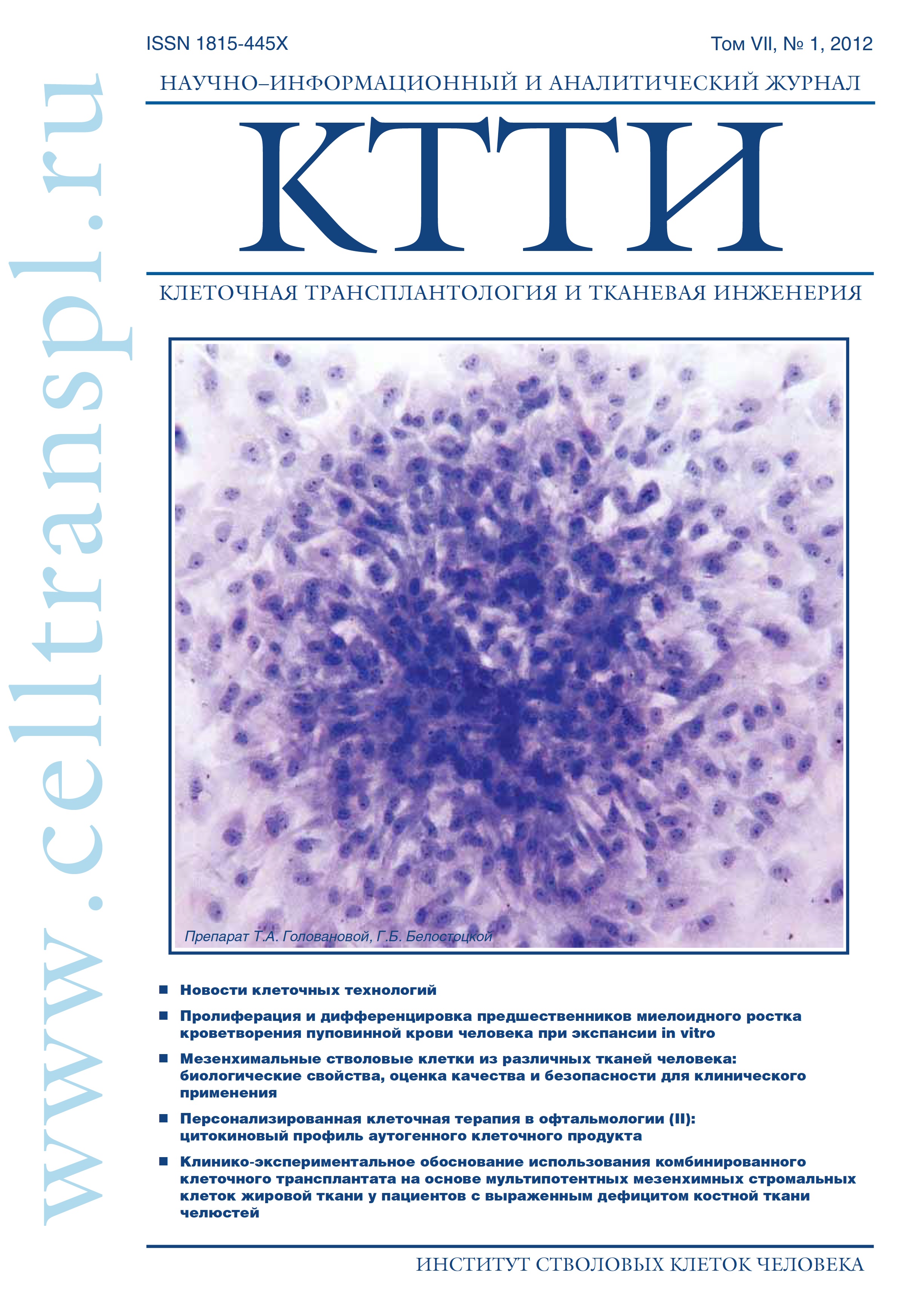
- Authors: Golovanova TA1, Belostotskaya GB1
-
Affiliations:
- Sechenov Institute of Evolutionary Physiology and Biochemistry of RAS, Saint PetersburgV.A. Almazov Federal Heart, Blood and Endocrinology Centre, Saint Petersburg
- Issue: Vol 7, No 1 (2012)
- Pages: 67-72
- Section: Articles
- Submitted: 11.01.2023
- Published: 15.03.2012
- URL: https://genescells.ru/2313-1829/article/view/121686
- DOI: https://doi.org/10.23868/gc121686
- ID: 121686

Cite item

Abstract
In parallel to the hypertrophy of major cardiac myocyte
population, we detected immunohistochemically the formation
of colonies consisting of small (dm = 6,20,5 m) resident
c-kit+ and Sca+ stem cells (SC) and Isl1+-positive cardiac
myocyte progenitors (CMP) in the primary culture of neonatal
rat cardiac myocytes. First contracting colonies (~1-2
clones per 100000 cells) were registered starting from 8th
day of culture. The cells of the colonies were capable of spontaneous
differentiation, demonstrating the maturation of contractile
machinery and Ca2+ responses caffeine (5 мМ) and
K+ (120 мМ). The full-scale development of electromechanical
coupling with typical for cardiac muscle Ca2+-induced Ca2+
release was obvious at 3 weeks of culture. At first, the local,
weak, spontaneous, asynchronous, and arrhythmic contractions
at a rate of 2-3 beats/min were registered. However,
with time the contractions became synchronous and involved
all cells of the colony with the rate of contractions being
58-60 beats/min at the end of the month. First contracting
clones comprised Isl1+ CMP, while c-kit+-colonies started to
contract 9-10 days later possibly owing to a more prolonged
period of proliferation.
Thus, we first demonstrated and characterized the
contracting colonies originating from SC and CMP when
those were co-cultivated with mature cardiac myocytes.
The process described in this study is akin to regenerative
cardiomyogenesis encompassing the pathway from resident
progenitor cell to the colony of mature contracting cardiac
myocytes. It follows, therefore, that contracting myocyte
colony is a suitable model for basic research, testing of drugs,
and the investigation of regenerative capacity of SC and CMP
aimed at future applications of resident progenitor cells in
cell-based treatment of cardiac injury.
About the authors
T A Golovanova
Sechenov Institute of Evolutionary Physiology and Biochemistry of RAS, Saint PetersburgV.A. Almazov Federal Heart, Blood and Endocrinology Centre, Saint PetersburgSechenov Institute of Evolutionary Physiology and Biochemistry of RAS, Saint PetersburgV.A. Almazov Federal Heart, Blood and Endocrinology Centre, Saint Petersburg
G B Belostotskaya
Sechenov Institute of Evolutionary Physiology and Biochemistry of RAS, Saint PetersburgV.A. Almazov Federal Heart, Blood and Endocrinology Centre, Saint PetersburgSechenov Institute of Evolutionary Physiology and Biochemistry of RAS, Saint PetersburgV.A. Almazov Federal Heart, Blood and Endocrinology Centre, Saint Petersburg
References
- Satin J., Itzhaki I., Rapoport S. et al. Calcium handling in human embryonic stem cell-derived cardiomyocytes. Stem Cells 2008; 26(8): 1961-72.
- Liu J., Lieu D.K., Siu C.W. et al. Facilitated maturation of Ca2+ handling properties of human embryonic stem cell-derived cardiomyocytes by calsequestrin expression. Am. J. Physiol. Cell Physiol. 2009; 297(1): 152-9.
- Asai Y., Tada M., Otsuji T.G. et al. Combination of functional cardiomyocytes derived from human stem cells and a highly-efficient microelectrode array system: an ideal hybrid model assay for drug development. Curr. Stem Cell Res. Ther. 2010; 5(3): 227-32.
- Kuzmenkin A., Liang H., Xu G. et al. Functional characterization of cardiomyocytes derived from murine induced pluripotent stem cells in vitro. FASEB 2009; 23(12): 4168-80.
- Beltrami A.P., Barlucchi L., Torella D. et al. Adult cardiac stem cells are multipotent and support myocardial regeneration. Cell 2003; 114: 763-76.
- Oh H., Bradfute S.B., Gallardo T.D. et al. Cardiac progenitor cells from adult myocardium: Homing, differentiation, and fusion after infarction. PNAS 2003; 100(21): 12313-18.
- Laugwitz K-L., Moretti A., Lam J. et al. Postnatal isl1+ cardioblasts enter fully differentiated cardiomyocyte lineages. Nature 2005; 433: 647-53.
- Behfar A., Crespo-Diaz R., Nelson T.J. et al. Stem cells: clinical trials results the end of the beginning or the beginning of the end?Cardiovasc. Hematol. Disord. Drug Targets. 2010; 10(3): 186-201.
- Malliaras K., Marban E. Cardiac cell therapy: where we've been, where we are, and where we should be headed. Br. Med. Bull. 2011; 98(1): 161-85.
- Голованова Т.А., Белостоцкая Г.Б. Способность миокарда крыс к самообновлению в экспериментах in vitro: пролиферативная активность неонатальных кардиомиоцитов. Клеточная транспланто- логия и тканевая инженерия 2011; VI (4): 66-70.
- Grynkiewicz G., Роenie M., Tsien R.Y. A new generation of Ca2+ indicators with greatly improved fluorescence properties. J. Biol. Chem. 1985; 260: 3440-50.
- Bers D.M. Excitation-contraction coupling and cardiac contractile force. 2d. Dordrecht, Boston, London: Kluwer Academic; 2001.
- Escobar A.L., Ribeiro-Costa R., Villalba-Galea C. et al. Developmental changes of intracellular Ca2+ transients in beating rat hearts. Am. J. Physiol. Heart. Circ. Physiol. 2004; 286: 971-8.
- Lieu D.K., Liu J., Siu C.W. et al. Absence of transverse tubules contributes to non-uniform Ca(2+) wavefronts in mouse and human embryonic stem cell-derived cardiomyocytes. Stem Cells Dev. 2009; 18(10): 1493-500.
- Satin J., Kehat I., Caspi O. et al. Mechanism of spontaneous excitability in human embryonic stem cell derived cardiomyocytes. J. Physiol. 2004; 559(Pt 2): 479-96.
Supplementary files
