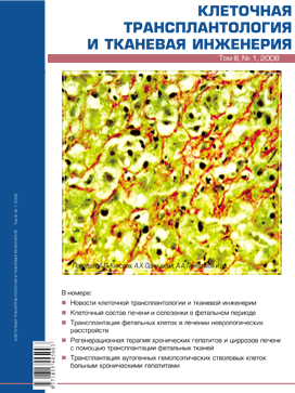
Transplantation of autologous hemopoietic stem cells to patients with chronic hepatitis
- Authors: Kiasov A.P.1, Odintsova A.K.2, Gumerova A.A.1, Gazizov I.M.1, Farrakhov A.Z.2, Kundakchyan G.G.1, Abdulkhakov S.R.1, Cheremina N.A.2, lilmaz T.S.1
-
Affiliations:
- Kazan State Medical University
- Republic Clinical Hospital of the Tatarstan Public Health Ministry
- Issue: Vol 3, No 1 (2008)
- Pages: 70-75
- Section: Clinical experience
- URL: https://genescells.ru/2313-1829/article/view/317444
- ID: 317444
Cite item
Abstract
The results of the clinic triai first phase of autotransplantation efficacy of hemopoietic stem cells to patients with chronic hepatitis in a severe hepatic fibrosis stage are given in the article. After hemopoietic precursors having been stimulated by "Neipogen" into peripheral blood, isolated hemopoietic stem cells were introduced into the celiac trunk. It provided the delivery of transplanted cells into the liver with both the arterial and venous blood flow. As the assessment criteria of transplantation efficacy the morphologic analysis results (the histologic activity index) and immunohistochemical staining (detection of proliferation activity, myofibroblasts activation and capillarization of sinusoids) were used together with clinical and biochemical values. The investigation results show that transplantation of autologous hemopoietic stem cells into the celiac trunk ofpatients with chronic hepatitis in the stage ofsevere fibrosis is a safe procedure. A month after transplantation the patient’s general condition and the functional tests indices improved, the necrosis and inflammation became less pronounced, qualitative structure alterations within the liver occured - that is hepatocytes proliferation decreased significantly, the number of myofibroblasts was reduced within the parenchyma that resulted in resolving of perisinusoidal fibrosis and restoration of the norma/ structure of sinusoid capillaries. The data obtained confirm that this method can be used to suppress the progression of hepatitis and fibrosis formation within the liver, as well as immunohistochemical methods provide reliable information on the efficacy of transplantation.
Full Text
About the authors
A. P. Kiasov
Kazan State Medical University
Author for correspondence.
Email: info@eco-vector.com
Deparment of Anatomy; Stem cell bank
Russian Federation, KazanA. Kh. Odintsova
Republic Clinical Hospital of the Tatarstan Public Health Ministry
Email: info@eco-vector.com
Russian Federation, Kazan
A. A. Gumerova
Kazan State Medical University
Email: info@eco-vector.com
Deparment of Anatomy
Russian Federation, KazanI. M. Gazizov
Kazan State Medical University
Email: info@eco-vector.com
Deparment of Anatomy; Stem cell bank
Russian Federation, KazanA. Z. Farrakhov
Republic Clinical Hospital of the Tatarstan Public Health Ministry
Email: info@eco-vector.com
Russian Federation, Kazan
G. G. Kundakchyan
Kazan State Medical University
Email: info@eco-vector.com
Stem cell bank
Russian Federation, KazanS. R. Abdulkhakov
Kazan State Medical University
Email: info@eco-vector.com
Deparment of Anatomy
Russian Federation, KazanN. A. Cheremina
Republic Clinical Hospital of the Tatarstan Public Health Ministry
Email: info@eco-vector.com
Russian Federation, Kazan
T. S. lilmaz
Kazan State Medical University
Email: info@eco-vector.com
Deparment of Anatomy; Stem cell bank
Russian Federation, KazanReferences
Supplementary files


