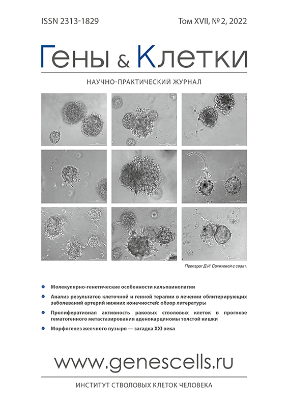
Angiogenic properties of glial progenitor cells derived from human induced pluripotent stem cells
- Authors: Salikhova D.I.1, Khaerdinova L.R.1, Makhnach O.V.1, Goldshtein D.V.1
-
Affiliations:
- N.P. Bochkov Research Centre for Medical Genetics
- Issue: Vol 17, No 2 (2022)
- Pages: 32-39
- Section: Original Study Articles
- URL: https://genescells.ru/2313-1829/article/view/133679
- DOI: https://doi.org/10.23868/202209005
- ID: 133679
Cite item
Abstract
Diseases associated with impaired blood supply to the brain ranks second term of mortality in the world, losing the place only to coronary heart disease. The incidence of this disease in the world remains high and increasing significantly with the age. The recent year’s special attention has been paid to the search for new methods of therapy for ischemic diseases, such as study of angiogenic properties of stem cells and their conditioned medium. The aim of this work is studying the angiogenic properties of glial progenitor cells derived from human induced pluripotent stem cells. The study was carried out by testing the proliferative activity, mobility, migration of endothelial cells line EA.hy926 under the influence of glial progenitor cells and their conditioned medium. Also the research was conducted by ability to formation of the tubular and capillary-like structure by cells line EA.hy926 by modeling angiogenesis in the basement membrane matrix in vitro.
The conditioned medium obtained by glial progenitor cells at concentrations of total protein 1 and 5 µg/ml has a positive influence on the proliferative activity and mobility of the endothelial cells line EA.hy926. At the same time it does not accelerate the formation of the primary tubular and capillary-like structure by the modeling angiogenesis in the basement membrane matrix in vitro. But glial progenitor cells contribute to the formation of tubular and capillary-like structure due to contact-dependent signaling between the two cell types. The primary formed tubular structure had a long processes and large branch points under co — cultivation with glial progenitor cells. Sprouting centers also had long and more convoluted processes and large cell clusters during the formation of a capillary-like structure. The glial progenitor cells and their conditioned medium had a positive effect on endothelial cell migration. This effect probably indicated by the production of substances by glial progenitor cells which was chemoattractants for endothelial cells line EA.hy926.
Full Text
About the authors
D. I. Salikhova
N.P. Bochkov Research Centre for Medical Genetics
Author for correspondence.
Email: diana_salikhova@bk.ru
Russian Federation, Moscow
L. R. Khaerdinova
N.P. Bochkov Research Centre for Medical Genetics
Email: diana_salikhova@bk.ru
Russian Federation, Moscow
O. V. Makhnach
N.P. Bochkov Research Centre for Medical Genetics
Email: diana_salikhova@bk.ru
Russian Federation, Moscow
D. V. Goldshtein
N.P. Bochkov Research Centre for Medical Genetics
Email: diana_salikhova@bk.ru
Russian Federation, Moscow
References
- Carmeliet P. Angiogenesis in life, disease and medicine. Nature 2005; 438(7070): 932–6.
- Darweesh S.R., Ayoub N.B., Nazzal S. Gold nanoparticles and angiogenesis: molecular mechanisms and biomedical applications. International Journal of Nanomedicine 2019; 14: 7643–63.
- Veith A.P., Henderson K., Spencer A. et al. Therapeutic Strategies for Enhancing Angiogenesis in Wound Healing. Advanced Drug Delivery Reviews 2019; 146: 97–125.
- Kuriakose D., Zhicheng X. Pathophysiology and Treatment of Stroke: Present Status and Future Perspectives. International Journal of Molecular Sciences 2020; 21(20): 7609–33.
- Ylä-Herttuala S., Rissanen T.T., Vajanto I. et al. Vascular Endothelial Growth Factors: Biology and Current Status of Clinical Applications in Cardiovascular Medicine. Journal of the American College of Cardiology 2007; 49(10): 1015–26.
- Рубина К.А., Семина Е.В., Дыйканов Д.Т. и др. Эффективность сочетанного использования плазмидных конструкций, содержащих гены hgf и ангиопоэтина-1, для восстановления кровотока в ишемизированных тканях. Гены и Клетки 2018; 13(1): 56–64. [Rubina K.A., Semina E.V., Dyikanov D.T. et al. The effectiveness of the combined use of plasmid constructs containing the hgf and angiopoietin-1 genes for restoring blood flow in ischemic tissues. Genes and Cells 2018; 13(1): 56–64].
- Nikfarjam S., Rezaie J., Zolbanin N.M. et al. Mesenchymal stem cell derived-exosomes: a modern approach in translational medicine. Journal of Translational Medicine 2020; 18(1): 1–21.
- Marcozzi C., Frattini A., Borgese M. et al. Paracrine effect of human adipose-derived stem cells on lymphatic endothelial cells. Regenerative Medicine 2020; 15(9): 2085–98.
- Макаревич П.И., Болдырева М.А., Дергилёв К.В. и др. Трансплантация клеточных пластов из мезенхимальных стромальных клеток жировой ткани эффективно индуцирует ангиогенез в ишемизированных скелетных мышцах. Гены и Клетки 2015; 10(3): 68–77. [Makarevich P.I., Boldyreva M.A., Dergilev K.V. et al. Transplantation of cell sheets from adipose-derived mesenchymal stromal cells effectively induces angiogenesis in ischemic skeletal muscle. Genes and Cells 2015; 10(3): 68–77].
- Huang Y., Wang L., He B. et al. Bone marrow mesenchymal stem cell-derived exosomes promote rotator cuff tendon-bone healing by promoting angiogenesis and regulating M1 macrophages in rats. Stem Cell Research & Therapy 2020; 11(1): 1–16.
- Hade M.D., Suire C.N., Suo Z. Mesenchymal Stem Cell-Derived Exosomes: Applications in Regenerative Medicine. Cells 2021; 10(8): 1959–2007.
- Yu F., Fu R., Liu L. et al. Leptin-Induced Angiogenesis of EA.Hy926 Endothelial Cells via the Akt and Wnt Signaling Pathways in vitro and in vivo. Frontiers in Pharmacology 2019; 10: 1275–90.
- Salikhova D., Bukharova T., Cherkashova E. et al. Therapeutic effects of hipsc-derived glial and neuronal progenitor cells-conditioned medium in experimental ischemic stroke in rats. International journal of molecular sciences 2021; 22(9): 4694–714.
- Arutyunyan I.V., Kananykhina E.Yu., Fatkhudinov T.Kh. et al. Angiogenic Potential of Multipotent Stromal Cells from the Umbilical Cord: an In Vitro Study. Bulletin of Experimental Biology and Medicine 2016; 161(1): 141–9.
- Konstantinou E.K., Notomi S., Kosmidou C. et al. Verteporfin-induced formation of protein cross-linked oligomers and high molecular weight complexes is mediated by light and leads to cell toxicity. Scientific reports 2017; 7(1): 1–11.
- Choi M., Lee H.S., Naidansaren P. et al. Proangiogenic features of Wharton's jelly-derived mesenchymal stromal/stem cells and their ability to form functional vessels. The International Journal of Biochemistry & Cell Biology 2013; 45(3): 560–70.
- Chen J., Liu Z., Hong M. et al. Proangiogenic compositions of microvesicles derived from human umbilical cord mesenchymal stem cells. Plos Оne 2014; 9(12): 1–16.
- Sun J., Shen H., Shao L. et al. HIF-1α overexpression in mesenchymal stem cell-derived exosomes mediates cardioprotection in myocardial infarction by enhanced angiogenesis. Stem Cell Research & Therapy 2020; 11(1): 1–13.
- Takeuchi R., Katagiri W., Endo S. et al. Exosomes from conditioned media of bone marrow-derived mesenchymal stem cells promote bone regeneration by enhancing angiogenesis. Plos Оne 2019; 14(11): 1–19.
- Walter M., Kohli N., Khan N. et al. Human mesenchymal stem cells stimulate EaHy926 endothelial cell migration: combined proteomic and in vitro analysis of the influence of donor-donor variability. Journal of Stem Cells & Regenerative Medicine 2015; 11(1): 18–24.
- Gong M., Yu B., Wang J. et al. Mesenchymal stem cells release exosomes that transfer miRNAs to endothelial cells and promote angiogenesis. Oncotarget 2017; 8(28): 45200–12.
Supplementary files


