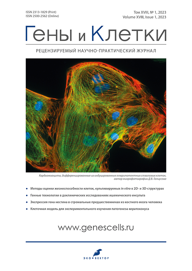
Vol 18, No 1 (2023)
Reviews
Methods for assessing the viability of cells cultured in vitro in 2D and 3D structures
Abstract
Over the past decades, cell viability tests have been an essential research tool in cell biology, tissue engineering, and regenerative medicine. Assessment of cell viability is mandatory in the production and quality control of cell products for biomedical applications.
Methods of viability assessment can be broadly classified according to the underlying mechanism and how the results obtained are evaluated. This article presents variants of the most commonly used tests and protocols for assessing cell viability. Their advantages and disadvantages are presented, which should be considered when planning experiments, e.g., when developing cell preparations for regenerative medicine.
The authors point out the main factors influencing the choice of viability assessment method: efficiency, speed, safety, reproducibility, sample integrity, compatibility with biomaterial, and cell line type. Finally, the authors discuss separately cell viability tests that can be applied not only to 2D cell structures but also to 3D cell structures, which have recently become widespread due to more accurate modeling of biological processes.
 5-21
5-21


Gene technologies in ischemic stroke preclinical studies
Abstract
Ischemic stroke is one of the leading death causes and disability worldwide. The review is devoted to analysis of gene technologies application achievements in ischemic stroke experimental study.
In experimental brain ischemia, genetic constructs effectiveness studies, genes, encoding predominantly neurotrophic and angiogenic factors delivery is actively pursued. Direct gene therapy has proven its effectiveness in experimental ischemic stroke. Genetic constructs delivering methods with target genes into the ischemic brain using cellular carriers has advantages because of combined action of genes and transplanted cells. Studies on ischemic stroke models with cellular carriers overexpressing various neurotrophic and angiogenic factors confirm the safety and effectiveness of this approach, which allows us to consider transgenes cell-mediated delivery as a stroke treatment promising method.
Another significant area of gene technologies application, which is also mentioned upon in the review, is related to opto- and chemogenetic methods, which allowed obtaining new data of ischemic stroke pathogenesis cellular and molecular mechanisms.
Three main criteria were used in the review: volume of infarction, capillary density and motor activity for effectiveness comparative assessment of direct administration, transgenes number and cell-mediated delivery.
 23-40
23-40


Original Study Articles
Tumor cell-free DNA detection-based liquid biopsy of plasma and bile in pancreatic ductal adenocarcinoma: a pilot study
Abstract
INTRODUCTION: Plasma liquid biopsy with tumor cell-free DNA detection is one of the most promising technologies in pancreatic ductal adenocarcinoma diagnosis. Due to low levels of this biomarker in plasma its detectability turns out to be lower than expected. However, some patients with this disease present with biliary obstruction, which enables collection of bile for tumor cell-free DNA analysis.
THE AIM: To comparison of diagnostic potential of cell-free tumor DNA detection-based liquid biopsy of plasma and bile in pancreatic ductal adenocarcinoma patients.
MATERIAL AND METHODS: The pilot study included 15 primary untreated pancreatic ductal adenocarcinoma patients with biliary obstruction. Cell-free DNA was isolated from 5 mL of plasma and 5 mL of bile. Tumor cell-free DNA detection was performed using digital droplet PCR with KRAS gene mutations analysis: G12A, G12C, G12D, G12R, G12S, G12V, G13D и Q61H (183A>C), Q61H (183A>T), Q61K, Q61L, Q61R. False-positive droplets threshold was established during the analysis of plasma cell-free DNA samples from 15 healthy volunteers.
RESULTS: Tumor cell-free DNA was detected in 13 out of 15 bile samples, whereas among paired plasma samples only 9 were positive. All plasma positive samples had a paired positive bile sample. Tumor cell-free DNA levels were statistically significantly higher in bile than in plasma: 538.0 (4.1–1960.0) vs. 4.4 (0–27.6) copies/mL (p=0.005).
CONCLUSION: Bile liquid biopsy in pancreatic ductal adenocarcinoma patients with biliary obstruction is a promising alternative to plasma analysis, due to higher concentrations of tumor cell-free DNA.
 41-51
41-51


Nestin gene expression in stromal precursor cells from the human bone marrow
Abstract
INTRODUCTION: The hierarchy of stromal progenitors from the bone marrow is poorly characterized; multipotent mesenchymal stromal cells and colony-forming units of fibroblasts are isolated in culture. Mesenchymal stem cells do not have a unique combination of surface antigens, making it difficult to obtain the pure population. The expression of the nestin gene is often used as a marker of these cells.
AIM: To evaluate the level of expression of the nestin gene in multipotent mesenchymal stromal cells and in colony-forming units of fibroblasts and to characterize the change in its expression during the transition from oligopotent progenitor cells to monopotent ones.
MATERIALS AND METHODS: Stromal progenitors were analyzed in bone marrow samples from 19 donors by standard methods. A total of 296 individual clones of fibroblast colony-forming units were obtained from the same bone marrow samples. The cells were analyzed for the ability to differentiate toward the adipogenic and osteogenic lineages. Relative expression level of nestin gene was analyzed in all cells.
RESULTS: Mean relative expression level of nestin did not differ significantly in multipotent mesenchymal stromal cells (0.41±0.13) and in the total population of colony-forming units of fibroblasts (0.24±0.05). In individual clones of colony-forming units of fibroblasts, nestin expression was not significantly higher than in the total population (0.31±0.04). When analyzing colony-forming units of fibroblasts differing in their differentiation potential, the highest expression of nestin was found in the group of monopotent osteogenic progenitors, while its expression was significantly lower in oligopotent progenitors.
CONCLUSION: Nestin gene expression in mesenchymal stromal progenitors from the bone marrow is not specific for mesenchymal stem cells and cannot be used as a unique marker of this cell type. According to our data, a high level of nestin expression rather identifies monopotent osteogenic progenitors.
 53-60
53-60


Biochemical indicators of sperm plasma and morphofunctional features of spermatozoa in HIV-infected men
Abstract
BACKGROUND: The problem of male infertility particularly affects men with HIV infections, as semen volume and sperm motility are reduced by highly active antiretroviral therapy.
AIM: To analyze the metabolic parameters of spermatozoa, spermograms and spermatozoa morphology in HIV-infected men.
MATERIALS AND METODS: The study included 47 patients (aged 25–46 years) who were under observation at the clinic of Professor M.A. Florova (Samara). 2 groups were formed: main (n=22) — HIV-infected patients who want to have children (with an undetectable viral load (40–50 copies/ml) and taking antiretroviral therapy); control (n=25) — clinically healthy men with one child. The material was taken and the ejaculate was examined according to standardized methods proposed by WHO experts.
RESULTS: In HIV-infected men, spermatozoa motility was low (the number of progressively motile spermatozoa was 28.50±3.72%), the spermatozoa concentration was two times lower compared to the control group of men, the morphological characteristics of spermatozoa were significantly worse than in the control group: most often pathology of the head (21.75±1.10%) and neck of the spermatozoon (22.30±1.18%) was detected, which negatively affects the fertilizing ability.
In HIV-infected men, changes in metabolic metabolism were noted: the activity of creatine phosphokinase in both sperm plasma (661.95±1.08 U/l) and blood plasma (76.90±1.09 U/l) was lower, than in the control group (844.25±0.13 and 79.50±1.37 U/l, respectively), the glucose concentration (8.07±1.14 mmol/l) was 2 times higher than in the control group (3.06±1.09 mmol/l), the concentration of calcium 6.53±0.01 and sodium (119.20±1.23 mol/l) slightly exceeded those in the group of healthy patients (5.55±0.08 and 116.85±0.01 mol/l, respectively). Electron microscopic analysis revealed fragmentation of sperm DNA: the highest percentage of sperm with fragmented DNA was found in men with HIV infection (more than 23%).
CONCLUSION: It was revealed that in vivo fertilization in HIV-infected men is impossible in most cases. The study forms the basis for a future comprehensive assessment of the state of reproductive function in HIV-infected men to assess their fertility and the need for assisted reproductive technologies.
 61-67
61-67


Сell model for experimental research in keratoconus pathogenesis
Abstract
INTRODUCTION: Certain mineral elements dismetabolism is known to play an important part in the pathogenesis of the corneal degenerative and dystrophic diseases, as metalloenzymes participate in connective tissues metabolism affecting their properties. Thus, in keratoconus corneal tissue is depleted in iron, copper and zinc, what could be the underlying cause of cornea biomechanical properties impairment. Keratoconus modeling is complicated and practically not reproducible in animals, therefore cell models elaboration is very much in demand. Definite mineral elements content must be reduced in order to simulate pathological changes specific for keratoconic corneas.
PURPOSE: The aim of this study was an elaboration of a cell model suitable for keratoconus pathogenesis research. This goal achieving involved solving the following tasks: 1) to develop a tissue-engineered system that mimic healthy corneal stroma; 2) to develop a technique for selective depletion of the nutrient medium by mineral elements involved in keratoconus pathogenesis; 3) to assess the possibility of the designed tissue-engineered system growth in the depleted nutrient medium.
MATERIAL AND METHODS: The study was carried out with the primary culture of human keratocytes, which were used to build tissue-engineered systems of three types: on silicone, on a membrane, without a carrier. Cell cultures morphology was evaluated by light and electron microscopy. The nutrient media were zinc depleted via decationization of fetal bovine serum using two types of ion-exchange resins; mineral elements concentrations were evaluated by means of inductively coupled plasma mass spectrometry.
RESULTS: The tissue system engineered without a carrier in form of cell sheet was chosen as the most convenient model. The decationization of the serum by means of Chelex 100 resin was shown to be a successful method for tenfold zinc concentration reduction in the nutrient medium. Keratocytes cultivation in the form of cell sheet on a zinc-depleted medium was successfully approved.
CONCLUSION: Elaborated tissue-engineered system could be considered as a model of the corneal stroma under specific for keratoconus conditions of zinc depletion.
 69-77
69-77


Cultivation of limbal stem cells on a biopolymer carrier (preliminary study)
Abstract
BACKGROUND: Limbal stem cell deficiency is a complicated pathology of ocular surface, which is not always helped by conservative methods of treatment and surgery is limited by available sources of tissue. Consequently, searching for new effective methods of its treatment is now gaining popularity. The most promising approach is transplantation of tissue-engineered constructs consisting of cultured limbal stem cells (LSCs) and variety of biopolymer carriers.
AIM: This study was performed to obtain and characterize a tissue-engineered constructs consisting of cultured LSCs and collagen membrane.
MATERIALS AND METODS: The study was performed at the Krasnov Research Institute at the Krasnov Research Institute of Eye Diseases and Koltzov Institute of Developmental Biology in cooperation with IMTEK Ltd. with a series of experiments. Two Chinchilla rabbits with an average weight of 3.5 kg and age of 6 months have been involved in this trial. LSCs were isolated and cultured in vitro from the healthy eye of rabbits using a method modified by the authors. The abtained cells were then cultured for 14 days and transplanted to the collagen membrane, which was then examined using immunohistochemical analysis.
RESULTS: The cells isolated from the biopsy were a mixture of fibroblast-type cells and cells with characteristics of LSC. They maintained high survivability, proliferativity, phenotype and stemness on the collagen carrier according to immunofluorescent study.
CONCLUSIONS: Thus, the abstained tissue-engineered constructs could be used for further transplantation to the affected eye with limbal stem cell deficiency under experimental conditions.
 79-88
79-88


