

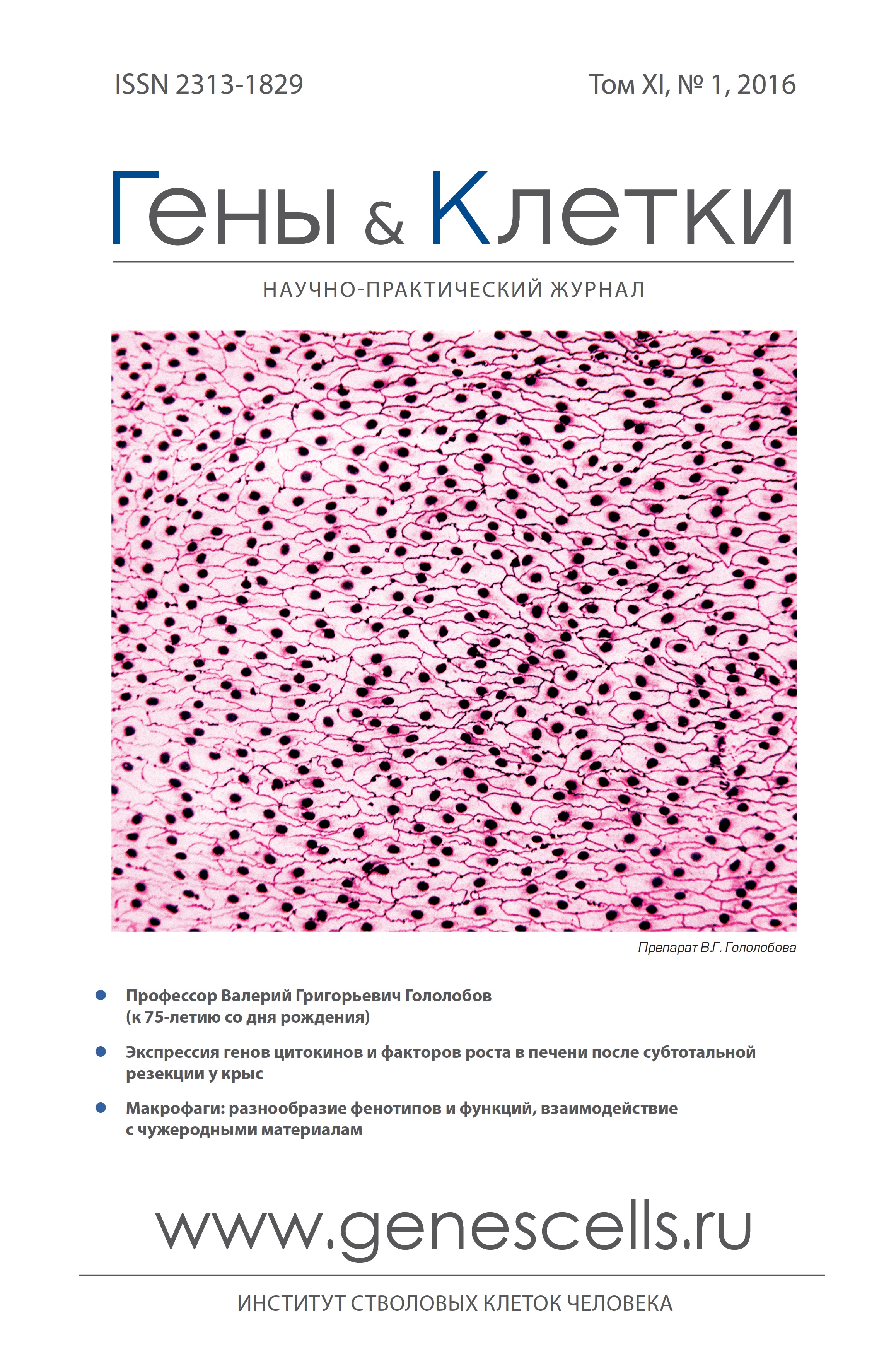
")


Vol 11, No 1 (2016)
- Year: 2016
- Articles: 12
- URL: https://genescells.ru/2313-1829/issue/view/6124
Articles
Professor Valery G. Gololobov (the 75th anniversary)
Genes & Cells. 2016;11(1):6-8
 6-8
6-8


Macrophages: diversity of phenotypes and functions, interaction with foreign materials
Abstract
According to “M1/M2” paradigm two distinct subsets of macrophages have been proposed - classically (M1) or alternatively (M2) activated macrophages that express different receptors, cytokines, chemokines, growth factors and effector molecules but recent evidence suggests that in respond to changed environmental stimuli they can demonstrate unique properties which do not allow to attribute them neither to M1 nor to M2 population. Macrophages play a pivotal role in foreign body reaction following installation of catheters, stents prosthesis, dental implants Macrophages englobe wear particles around joint prosthesis initiating an inflammation in periprosthetic tissues аnd osteolysis, control fibroproliferation and formation of the fibrous capsule surrounding foreign bodies A brief overview of events leading to migration, adhesion and activation of macrophages, and analysis of their functional characteristics on different surfaces including biodegradable and non-biodegradable materials in vivo and in vitro are presented
Genes & Cells. 2016;11(1):9-17
9-17
Encapsulation of cells and tissues of the pancreas: problems and ways of their overcoming
Abstract
Despite advances in treatment the patients suffering from diabetes mellitus type 1 have a lifetime shorter the average in population. This is defined primarily by the lack of sufficient glycemic control in these patients The active researches investigating the safety and efficacy of the grafting materials have been carried out in the last decades The review presents modern data on the use of pancreas cells and tissues encapsulation as a possible method for treatment of diabetes type 1 The main problems of the capsules application and possible ways to overcome them were described
Genes & Cells. 2016;11(1):18-23
18-23
INSULIN-PRODUCING CELLS IN THE TREATMENT OF INSULIN-DEPENDENT DIABETES MELLITUS
Abstract
An effective treatment for insulin-dependent diabetes mellitus (DM), which provides an alternative to hormone replacement therapy, is transplantation of insulin-producing cells (IPCs). Donor β-cells are transplanted both in the form of a complete pancreas, or in the form of isolated islets of Langerhans. However, the application of this method is limited due to the lack of donor material and the need for lifelong immunosuppressive therapy that has a detrimental impact on the weakened DM patient's body. An alternative method of obtaining IPCs is to differentiate stem or progenitor cells. Pancreatic differentiation capability has been demonstrated for various types of stem cells Currently, induced pluripotent stem cell IPSC) are considered to be the most promising source of IPCs, including those obtained from mature cells of the patients themselves Firstly, such IPCs can be gained in unlimited quantities. Secondly, in the case of autologous transplantation they are least exposed to the recipient body's immune attack, thereby making it possible to completely discard immunosuppressive therapy. IPSCs introduction into clinical practice is hindered by the fact that they provoke the formation of teratomas in the recipient>s body. Moreover, they retain this ability even after differentiation because of a number of undifferentiated cells preserved in the population This review focuses on contemporary protocols for obtaining IPCs from IPSCs. These protocols mimic β-cells formation stages during embryonic development. The review also covers the application of IPC immuno-isolating containers for transplantation. Their semipermeable walls, on the one hand, protect the transplant from the recipient>s immune system, and on the other hand, they suppress the risk of the transplant causing tumor formation. in addition, attention will be paid to the use of IPCs derived from IPSCs as a model object for studying the processes occurring in β-cells at normal circumstances as well as during DM.
Genes & Cells. 2016;11(1):24-34
24-34
Adipose-derived stromal vascular fraction as an alternative source of cells for the regenerative medicine
Abstract
The adipose tissue is considered as the most convenient and abundant source of cells for the regenerative medicine. The number of progenitor cells in the adipose tissue significantly exceeds their amount in the bone marrow and other tissues. Therefore, adipose-derived stromal vascular fraction comprising distinct populations of stem and progenitor cells can be relatively easily isolated from lipoaspirates and may then be used in various pathological conditions. However, the profile of this cell fraction with a significant therapeutic potential remains unclear, and there are no standardized protocols for its isolation and evaluation. in this article, we reviewed the data on the potential use of adipose-derived stromal vascular fraction in the regenerative medicine. We described the main historical milestones and performed a comprehensive analysis of the sources of adipose-derived stromal vascular fraction, techniques of its isolation, features, immunophenotype and differentiation pathways
Genes & Cells. 2016;11(1):35-42
35-42
Prospects for tissue engineered bile duct
Abstract
Intraoperative bile duct injures requiring its repair observed in 0. 05-2. 7% of patients, who underwent cholecystectomy due to cholelithiasis Lots of patients require reconstructive bile duct surgery given that cholecystectomy is the second most common surgery in the abdominal region, and more than 1 mln operations are made all over the world per year. Previously stitching of the crossed bile duct edges was used, but in most cases this entailed the bile duct stricture and disturbance of the bile outflow. At present, the standard surgery includes suturing of the duct with small intestine, but such a reconstruction, in turn, can lead to liver abscess, biliary cirrhosis and increased risk of cholangiocarcinoma. in this review, we consider the possibility of creating fragments of tissue-engineered bile duct that involves the use multilayer tissue-engineered structures consisting of a composite matrix, cells and signaling molecules that stimulate local proliferation and neovascularization.
Genes & Cells. 2016;11(1):43-47
43-47
The effects of co-culture duration of cord blood cells with adipose tissue-derived stromal cells on hematopoietic precursors> amplification
Abstract
Umbilical cord blood is considered as a valuable source of hematopoietic stem and progenitor cells (CB-HSPCs). The number of latter may be significantly enriched with ex vivo expansion. Thus, the optimization of culture conditions is essential for in vitro manipulations. Recently we have demonstrated that CB-HSPCs may be separated from unmanipulated CB nucleated cells through the adhesion to adipose tissue-derived MSCs. Further coculture was resulted in raizing of new polulation of floating CB-HSPCs significantly ehriched in primitive progentors The goal of this study was to optimize above mentioned protocol To determine the optimal conditions for adhesion and multiplication of CB-HSPCs, nucleated CB cells were co-cultured on adipose-tissue MSC layer for short (1-3 hours) and long-term (24-72 hours) duration Unattached cells were removed, adherent CB-HSPCs were further cultured for 72 hours, resulted in formation of floating population of CB-HSPCs. in each time point the number of attached CB-HSPCs, newly formed floating CB-HSPCs, CD34+ cells and CFUs among latter was examined. After 72 hours of nucleated CB cells co-culture, the number of adherent CD34+ cells peaked and was over than 70% of total CD34+ cells among nucleated CB cell samples Proposed experimental design has provided 4-fold enrichment of primitive CD34+ and 6-fold of CFUs number among newly formed HSPCs. BFU-Es comprised 80-90% of total CFUs regardless of time of nucleated CB cells coculture. Thus, 3 days of nucleated CB cells/adipose tissue-derived mesenchymal stromal cells co-culture provided peak of CD34+ cells' adhesion, amplification of latter resulted in rising of population maximally enriched both with undifferentiated and committed hematopoietic precursors
Genes & Cells. 2016;11(1):48-53
48-53
Hepato-specific small-dispersioned matrix as the important component of implanted cell-engineering designs for an auxiliary liver
Abstract
Biocompatible and biodegraded matrixes have already framed by the modern biomedical technologies however working out and application of tissue-specific matrixes remains an actual problem of the modern tissue engineering Aim of this work is to show that the technique, which is proposed by the authors for producing of small-dispersioned matrices of decellularization liver (DCL), is good for the making of cell-engineering designs of auxiliary liver The paper presents the technology of producing the small-dispersioned matrix of DCL and the results of using the light, phase- contrast and electron microscopy to characterize the biological properties of the made matrix The hepatospecific properties of the matrix have been studied by using a quantitative evaluation of the MTT-test results on the 5th day of separate cultivation on this matrix 4 types of cells (HepG2, renal epithelial cells, bone marrow MSCs and liver cells from allogeneic donors). On photographs of microscopic examination of the matrix particles of DCL it was seen their porous structure, on the surface of which the preserved conglomerates of native extracellular matrix molecules were presented At comparative study of adhesive properties of the Cytodex-3 matrix particles and the small-dispersioned matrix of DCL it was found out that both matrices had the ability to adhere different cell types, but the matrix of DCL had the ability to preserve of hepato-specific activity significantly expressed. Keeping of biocompatibility and hepato-specific properties by small-dipersioned matrices of DCL, produced on the proposed technology, allows to recommend them for the making of implantable cell-engineering designs of auxiliary liver.
Genes & Cells. 2016;11(1):54-60
54-60
Cytokines and growth factors genes expression after subtotal liver resection in rats
Abstract
Liver regeneration after 70% resection is one of the most studied models of tissue regeneration Small residual liver volume after massive hepatectomy or partial liver transplantation is a major cause of subsequent liver dysfunction known in oncology and transplantology as small-for-size syndrome The aim of this study was to evaluate the influence of 80% liver resection on cytokines and growth gens expression in rat Male Wistar rats were subjected to 80% and sacrificed at different times after surgery. Untreated animals served as controls Serum and liver samples were obtained to investigate liver function, cytokines and growth factors genes expression: il1b, il6, il10, tnfa, tweak, mmp9, fgf2, tgfb, hgf, vegf, sdfa, ang with quantitative RT-PCR. It was revealed two phases increased expression of genes that correspond to the first and second peak hepatocytes proliferation The first phase was characterized by increased expression mmp9, il6, il10, fgf2, tgfb. The second phase was characterized by increased expression of hgf, tnfa, tweak, il1b. Furthermore, we have found increased expression of the transcription factor SOX9 gene, and the gene encoding TWEAK, indicating a possible role for resident progenitor cells in the regenerating liver of rats after subtotal resection In our opinion the two phases of increased gene expression during liver regeneration in rats after subtotal resection are associated with two waves of hepatocyte proliferation, and the presence of growth factors reserves in the liver.
Genes & Cells. 2016;11(1):61-67
61-67
Histological features of the lipograft with platelet-rich plasma after subcutaneous transplantation in vivo
Abstract
Autologous adipose tissue transplantation is one of the most common methods of soft tissue volume and shape correction However, the effect of fat grafting is short due to the low survival rate of a fat graft This study was designed to evaluate the influence of the platelet-rich plasma (PRP) on the dynamics of involutive changes of the fat graft in an experimental model with subcutaneous injection in the rabbit ear shell Each animal (n = 9) underwent subcutaneous autologous fat grafting: in the left ear - without PRP, in the right one - mixed with PRP We performed the histological analysis of the materials estimating the number of fat elements, fibrosis processes, and severity of the macrophage-histiocytic reaction (identification of CD163+-cells) in 1, 2, 4, 8, 36 weeks after surgery We found that in the early stages of observation, up to 4 weeks, the signs of fibrosis (the thickness of the connective tissue capsule, interlobular and interadipocyte fibrous septums) were less pronounced in the case of PRP However, further histologic characteristic became similar in the both groups Thus, PRP can have a positive impact on the autologous fat graft survival, but more research is needed to select the optimal PRP processing protocol for autologous fat graft and further analysis of the causes, mechanisms and symptoms of the transplanted fat tissue involution
Genes & Cells. 2016;11(1):70-74
70-74
Problems of biomedical technologies legal regulation in Russia and abroad
Abstract
The work provides an overview of contemporary normative acts of the Russian Federation devoted to specific aspects of biomedicine: reproductive technologies, genetic engineering activity and genomic testing, treatment of embryos and fetal tissue. We analyze the draft federal law «On biomedical cell products», passed by the State Duma on 21 April 2015 in the first reading. The drawbacks of the project, presented the main points of view in terms of its legal regulation, formulated by a professional society of lawyers and experts in the field of cell technology. An overseas and international experience. In particular, the general provisions of the legislation of France, Germany, the Convention on Human Rights and Biomedicine, developed by the Council of Europe and adopted in Oviedo in 1997, the European Directive (2004/23 / EC) for the establishment of standards of quality and safety of organ donation, collection, testing, handling, storage and distribution of human tissues and cells We formulate a general conclusion on the need for the base of the Federal Law «On Biomedicine», which could only be resolved key issues that are at the peak of the scientific discussion of the professional community, affecting basic human rights All technological issues need to be resolved at the level of subordinate legislation
Genes & Cells. 2016;11(1):75-81
75-81
Ethical and scientific aspects of human embryonic material research: the Great Britain regulations
Abstract
We analyze the ethical and scientific issues of human embryonic specimen and its utilization for research purposes. Human embryonic specimens are the source of stem cells which are the foundation for all organs and tissue formation. The analysis of human embryonic specimens promises to obtain the essential knowledge about human development and reveal the causes of human development pathology. Ethical and scientific problems of human embryonic specimens donation and research are debated here We review the impact that human embryonic specimens impose on scientific research, usage restrictions, risk assessment, research personnel and donor protection in view of the British, and other European publications, case reports, legislation, regulations and guidelines This work is focused on the discussion of different aspects of donation and usage of human embryonic specimens obtained after the elective medical termination of pregnancy We discuss suggestions for improvement of the ethical and scientific regulations for the work with human embryonic donor material in Russian Federation.
Genes & Cells. 2016;11(1):82-89
82-89
СМИ зарегистрировано Федеральной службой по надзору в сфере связи, информационных технологий и массовых коммуникаций (Роскомнадзор).
Регистрационный номер и дата принятия решения о регистрации СМИ:
Регистрационный номер и дата принятия решения о регистрации СМИ: