

")


Optimal decellularization of rat hearts and diaphragms and morphological evaluation
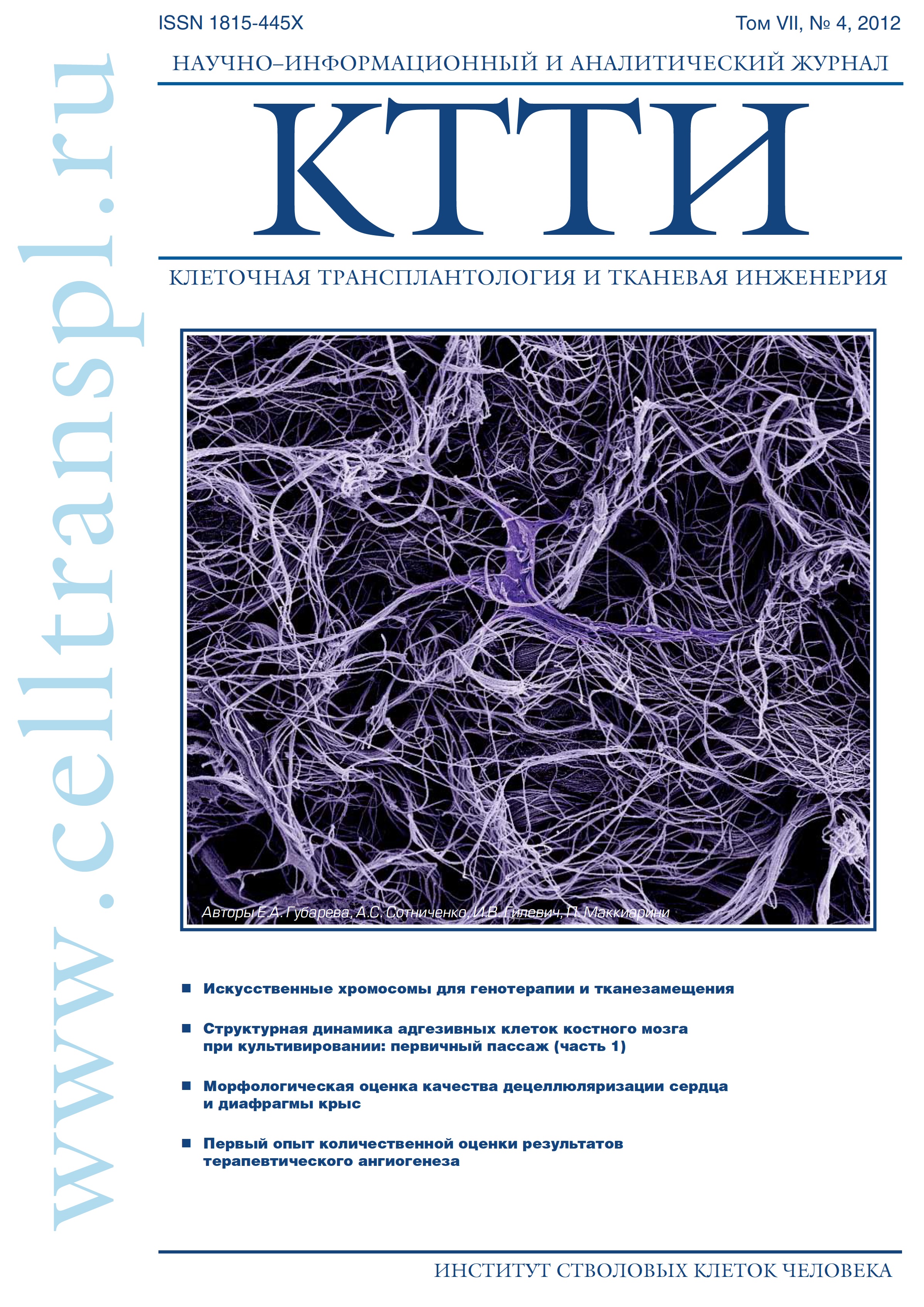
- Authors: Gubareva E.A.1, Sotnichenko A.S1, Gilevich I.V1, Macchiarini P.1,2
-
Affiliations:
- International Research, Clinical and Education Center of Regenerative Medicine, Kuban State Medical University, Krasnodar, Russia
- Advanced Center for Translational Regenerative Medicine (ACTREM), Karolinska Institutet, Stockholm, Sweden
- Issue: Vol 7, No 4 (2012)
- Pages: 38-45
- Section: Articles
- URL: https://genescells.ru/2313-1829/article/view/121571
- DOI: https://doi.org/10.23868/gc121571
- ID: 121571

Cite item

Abstract
Tissue engineering involves the design, evaluation, modification and maintenance of living cells or tissues embedded in biological (natural) or artificial scaffolds. Biological scaffolds need to be decellularized to become completely non-immunogenic while preserving the tissue structure and extracellular matrix. The primary goals of the present paper were to investigate and optimize different decellularization protocols of rats heart and diaphragm and establish the most reliable technique (paraffin vs. cryosections) to evaluate the morphology of the decellularized tissues. Hearts and diaphragm were decellularized with detergent-enzymatic based protocols, including deoxycholate and DNAse. Compared to published decellularization protocols, our was able to reduce exposure time (for heart: up to 3 hours — deoxycholate and 1 hour — DNAse; for diaphragm: up to 6 hours — deoxycholate and 2 hour — DNAse) of detergents and length (up to 24 hours). Results of morfological studies showed the absence of cells and preservation of the extracellular matrix. DNA quantification showed that about 81% and 74% of heart and diaphragm nuclear material, respectively, was removed by the decellularization process. Our results suggest that the investigated decellularization protocol was superior to others in removing DNA content and preserving the ECM of rats hearts and diaphragms.
Keywords
Full Text
About the authors
E. A. Gubareva
International Research, Clinical and Education Center of Regenerative Medicine, Kuban State Medical University, Krasnodar, Russia
A. S Sotnichenko
International Research, Clinical and Education Center of Regenerative Medicine, Kuban State Medical University, Krasnodar, Russia
I. V Gilevich
International Research, Clinical and Education Center of Regenerative Medicine, Kuban State Medical University, Krasnodar, Russia
P. Macchiarini
International Research, Clinical and Education Center of Regenerative Medicine, Kuban State Medical University, Krasnodar, Russia; Advanced Center for Translational Regenerative Medicine (ACTREM), Karolinska Institutet, Stockholm, Sweden
References
- Мировая статистика здравоохранения. ВОЗ. 2011. http:// www.who.int/gho/publications/world_health_statistics.
- Fuchs J.R., Nasseri B.A., Vacanti J.P. Tissue engineering: a 21st century solution to surgical reconstruction. Ann. Thorac. Surg. 2001; 72: 577-91.
- Mclntire L.V., Greisler H.P., Griffith L. et al., WTEC Panel Report on Tissue Engineering Research. Final report. Baltimore, MD: International Technology Research Institute. 2002 Jan.
- Langer R., Vacanti J.P. Tissue engineering: the design and fabrication of living replacement devices for surgical reconstruction and transplantation. Science.1993; 260: 920-6.
- Skalak R., Fox C., editors. NSF Workshop, UCLA Symposia on Molecular and Cellular Biology. 1988.
- Amulya S. Tissue engineering: Present concepts and strategies. Journal of Indian Association of Pediatric Surgery. 2005; 10: 14-9.
- Atala A. Tissue engineering, stem cells and cloning: current concepts and changing trends Expert opinion on biological therapy. 2005; 5: 879.
- Badylak S.F., Taylor D., Uygun K. Whole-Organ Tissue Engineering: Decellularization and Recellularization of Three-Dimensional Matrix Scaffolds, Annu. Rev. Biomed. 2011; 13: 27-53.
- Ott H., Taylor D., inventors; Regents of the University of Minnesota, assignee. Decellularization and recellularization of organs and tissues. US patent 20090202977. 2009 Aug 13.
- Conconi M.T., De Coppi P., Bellini S. et al. Homologous muscle acellular matrix seeded with autologous myoblasts as a tissue- engineering approach to abdominal wall-defect repair. Biomaterials. 2005; 26: 2567-74.
- Macchiarini P., Jungebluth P., Go T. et al. Clinical transplantation of a tissue-engineered airway. Lancet. 2008; 372: 2023-30.
- Andrew P. P., England K.A., Matson A.M. et al. Development of a decellularized lung bioreactor system for bioengineering the lung: the matrix reloaded. Tissue engineering. Part A. 2010; 16: 2581-91.
- Badylak S.F. The extracellular matrix as a biologic scaffold material. Biomaterials. 2007; 28: 3587-93.
- http://www.nanodrop.com
- http://www.qiagen.com
- Nagata S., Hanayama R., Kawane K. Autoimmunity and the clearance of dead cells. Cell. 2010; 140(5): 619-30.
- Zhang Q., Raoof M., Chen Y. et al. Circulating mitochondrial DAMPs cause inflammatory responses to injury. Nature 2010; 464(7285): 104-7.
- Brown B.N., Barnes C.A., Kasick R.T. et al. Surface characterization of extracellular matrix scaffolds. Beer-Stolz D. Biomaterials. 2010; 31: 428-37.
- Brown B.N., Valentin J.E., Stewart-Akers A.M. et al. Macrophage phenotype and remodeling outcomes in response to biologic scaffolds with and without a cellular component. Biomaterials. 2009; 30(8): 1482-91.
- Zheng M.H., Chen J., Kirilak Y. et al. Porcine small intestine submucosa (SlS) is not an acellular collagenous matrix and contains porcine DNA: possible implications in human implantation. J Biomed. Mater. Res. 2005; 73(1): 61-7.
- Ahn S.J., Costa J., Emanuel J.R. PicoGreen quantitation of DNA: effective evaluation of samples pre- or post-PCR. Nucleic Acids Res. 1996; 24(13): 2623-5.
Supplementary files
